There are a variety of imaging technologies that can be used to look at a brain injury or a concussion, and are used in unique ways to address and review specific concerns with a head injury. These imaging technologies each have their place and use in addressing and understanding not only a brain injury and concussions but many injuries that may occur throughout our bodies. It is important to know that not all imaging technologies are alike or able to clear detect and diagnose a concussion.
CT Scan or CAT Scan – Computerized Axial Tomography Scan for Concussion
A CT Scan is essentially a 3D X-ray machine. When used to look at the brain, it technically looks at the structure of the brain. When used with contrast, it helps to increase the visibility of the images. A CT scan will send a series of narrow beams throughout the location being scanned to produce more details in the images than a standard x-ray. A CT scan is able to show when an organ has been torn or altered structurally from an injury.
SPECT Scan – Single Photon Emission Computed Tomography Scan for Concussion
This scan is an advanced form of a CT scan. Using a radioactive substance, it produces 3D images of the area being scanned. This scan can show varying activity levels in the brain. This type of scan can be helpful in seeing what areas are less active, but they do not address functioning abilities where activity is happening.
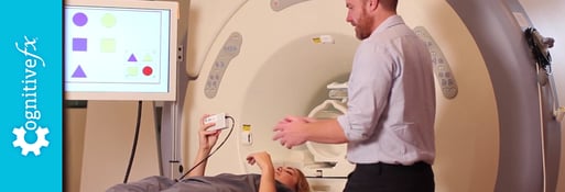
MRI – Magnetic Resonance Imaging for Concussion
An MRI scan uses a strong magnetic field and radio waves to create detailed images of organs and tissues within the body. This technology is noninvasive and painless. A standard MRI does not use any ionizing radiation which has the potential to harm an individual. MRI technology gives medical professionals the ability to see a detailed cross sectional image of internal images. This imaging looks at structural injuries. Typically, the patient is laying in a relaxed state of being while getting an MRI.
PET Scan– Position Emission Tomography Scan for Concussion
Using radiation or nuclear medicine imaging, this scan produces 3D images of functional processes within the human body. These types of scans typically address specific conditions and how the condition is developing. As a patient breaks down the radioactive medicine, medical professionals can see how the body is functioning throughout the breakdown process.
The largest difference between a PET scan, CT scan, MRI scan, and a SPECT scan, is that a PET scan can address and understand brain functioning. The other scanning technologies solely look at structural changes or injuries as seen in the other scan types. The downside to this scan is that it is invasive and because of the radioactive medicine it brings along its own set of risks. Because of this, pregnant women cannot be scanned due to radiation risk with the baby.
fMRI – functional Magnetic Resonance Imaging for Concussion
This form of MRI technology measures brain activity by detecting changes associated with blood flow. We know that blood flow and neuronal activity are coupled together, making it vital to understanding this technology. This technology has the ability to look at functioning in the brain when it comes to neurons and their firing capabilities.
Often times, a patient is at rest in an fMRI scan, and is not participating in cognitive activities. There are some limitations as to who can be scanned because of the powerful magnet. Those who have materials in their body that are magnetic cannot be scanned; i.e. pacemakers, certain types of metal plates, etc.
fNCI – functional NeuroCognitive Imaging for Concussion
An adapted form of fMRI technology, this type of scan looks at blood flow and neuronal activation while a patient participates in cognitive tests, similar to those found in a paper pencil test. fNCI detects changes in neural activation while a patient is performing a cognitive test, providing a three dimensional map of brain regions that are used to perform the test.
Because the fNCI scan measures brain function rather than solely giving an image of brain structure, it has the potential to be a very powerful diagnostic and assessment tool. This is especially important for more subtle pathologies, such as mild TBI or mild cognitive impairment, where standard brain imaging rarely detects abnormalities in visible tissue. This gives physicians the ability to look at about 100 distinct brain regions, and look for neuronal biomarkers, or trending levels of dysregulation in the brain.
This scan for concussion, created by Notus Neuropsychological Imaging, is one of the most advanced scans available today. To understand what a normal functioning brain looked like, Notus NI scanned hundreds of individuals over many years. By directly assessing brain activation, clinicians find that fNCI provides more information about brain function in 24 minutes of patient testing than 6-10 hours of traditional neuropsychological testing. Because of this reliability, the fNCI scan is becoming the new gold standard for making important clinical decisions, such as prescribing treatment protocols, assessing treatment outcomes, and determining return-to-play readiness.
Overall, these different imaging technologies are used in hospitals and clinics all around the world to understand the impacts of injury.