POTS and Ehlers-Danlos Syndrome: Why They Travel Together, and What That Means for Your Care
If you have postural orthostatic tachycardia syndrome (POTS) and you have been told more than once that you are unusually flexible, the two are connected. Up to 61% of pediatric POTS patients also meet criteria for Ehlers-Danlos syndrome (EDS) or hypermobility spectrum disorder (HSD), and roughly 78% of EDS patients show signs of autonomic dysfunction. The two conditions share a likely root in connective tissue biology, and recognizing the overlap changes diagnosis, changes treatment, and changes what realistic recovery looks like.
This guide walks through the connection in plain language for patients who are already living with one diagnosis and trying to understand the other. It is also written for clinicians who want a single reference point on the comorbidity. The clinical details are sourced from peer-reviewed literature published between 2015 and 2026, with citations linked at the end of each section.
What is POTS, in one paragraph?
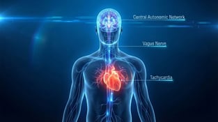
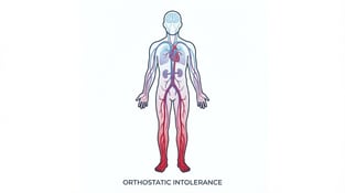
POTS is a form of orthostatic intolerance defined by a sustained heart rate increase of 30 beats per minute or more (40 bpm in patients under 20 years old) within 10 minutes of standing or head-up tilt, in the absence of orthostatic hypotension. The hallmark symptoms include lightheadedness, palpitations, fatigue, brain fog, exercise intolerance, gastrointestinal complaints, headache, and tremulousness on standing. It affects roughly 0.2% to 1% of people in developed countries, predominantly women between 15 and 50 years old, and typically begins after a triggering event such as a viral illness, surgery, trauma, pregnancy, or vaccination.
What is Ehlers-Danlos syndrome?
The Ehlers-Danlos syndromes are a group of 13 heritable connective tissue disorders, defined by the 2017 International Classification of the Ehlers-Danlos Syndromes published by Malfait and colleagues. Each subtype is characterized by some combination of joint hypermobility, skin involvement, and tissue fragility, and 12 of the 13 subtypes have identified causative genes. The most common subtype by far is hypermobile EDS (hEDS), which accounts for roughly 90% of all EDS cases and remains the only subtype without a confirmed genetic test.
Patients who have symptomatic joint hypermobility but do not meet the strict 2017 hEDS diagnostic criteria are now classified as having a hypermobility spectrum disorder (HSD). HSD is divided into four categories based on whether the hypermobility is generalized, peripheral, localized, or historical. Combined, EDS and HSD affect roughly 1 in 500 people, with HSD being more common than EDS.
The 2017 criteria are currently being revised. The Ehlers-Danlos Society's "Road to 2026" initiative, which includes the HEDGE genetic study of more than 1,000 hEDS patients sequenced at the Broad Institute, is scheduled to publish a new classification framework on December 1, 2026 in the American Journal of Medical Genetics. As of late 2025, no single causative gene for hEDS has been identified, and the working model points to multiple rare variants with complex interactions.
References for this section: Malfait F et al. The 2017 international classification of the Ehlers-Danlos syndromes. Am J Med Genet C Semin Med Genet. 2017;175(1):8-26. DOI Mehta D et al. Headache disorders in patients with Ehlers-Danlos syndromes and hypermobility spectrum disorders. Front Neurol. 2024;15:1460352. DOI Cutsforth-Gregory JK, Sandroni P. Clinical neurophysiology of postural tachycardia syndrome. Handb Clin Neurol. 2019;161:429-445. DOI
How often do POTS and EDS actually occur together?
More often than chance, and the numbers are striking in both directions.
Looking at POTS populations:
- Boris and Bernadzikowski (2020) evaluated 362 pediatric POTS patients. 22.7% met criteria for EDS and 39.0% met criteria for HSD. That is roughly 6 out of every 10 pediatric POTS patients carrying some form of joint hypermobility diagnosis. The POTS-plus-EDS group also had earlier symptom onset (median 12.1 years versus 13.5 years) and longer symptom duration before diagnosis.
- Miglis and colleagues (2017) compared 20 POTS-plus-hEDS adults to 20 POTS-only adults. The hypermobile group had roughly twice the outpatient visit count (21 versus 10), more prescription medications (8 versus 5.5), and a much higher rate of pain physician involvement (70% versus 25%), even though their autonomic test results were broadly similar.
- Mudie et al. (2024) studied 815 ME/CFS registry participants. 15.5% had joint hypermobility, and those with concurrent hypermobility and EDS had significantly worse health-related quality of life and a higher symptom burden across autonomic, neurocognitive, headache, gut, and musculoskeletal domains.
Looking at EDS populations:
- Up to 78% of EDS patients show some form of dysautonomia, with POTS the most commonly recognized form.
- Celletti and colleagues (2020) found POTS in 48% of 102 adults with hEDS or HSD using multimodal autonomic testing, with orthostatic intolerance in another 25.5%.
- Tai and colleagues (2020) surveyed 616 HSD/hEDS subjects through Ehlers-Danlos Support UK. 37.5% reported a doctor-confirmed POTS diagnosis, and that group had significantly worse gastrointestinal symptoms and a higher prevalence of chronic fatigue syndrome.
- Peebles and colleagues (2022) found POTS in 43% of women with generalized HSD on head-up tilt testing, compared to 7% of women with hEDS and 7% of healthy controls, although results varied depending on whether active stand or head-up tilt was used.
The practical takeaway is that any thorough POTS workup should include hypermobility screening, and any EDS or HSD workup should include orthostatic vital sign measurement. The two conditions miss each other constantly in standard practice, and both diagnoses tend to take years longer than they should.
References for this section: Boris JR, Bernadzikowski T. Prevalence of joint hypermobility syndromes in pediatric postural orthostatic tachycardia syndrome. Auton Neurosci. 2020;231:102770. DOI Miglis MG et al. Postural tachycardia in hypermobile Ehlers-Danlos syndrome: A distinct subtype? Auton Neurosci. 2017;208:146-149. DOI Mudie K et al. Do people with ME/CFS and joint hypermobility represent a disease subgroup? Front Neurol. 2024;15:1324879. DOI Celletti C et al. A new insight on postural tachycardia syndrome in 102 adults with hEDS/HSD. Monaldi Arch Chest Dis. 2020;90(2). DOI Tai FWD et al. Functional gastrointestinal disorders are increased in joint hypermobility-related disorders with concomitant POTS. Neurogastroenterol Motil. 2020;32(12):e13975. DOI Peebles KC et al. The prevalence and impact of orthostatic intolerance in young women across the hypermobility spectrum. Am J Med Genet A. 2022;188(6):1761-1776. DOI
Why do POTS and EDS occur together? What is the actual mechanism?
There is no single cause. Current research points to at least five overlapping mechanisms, and individual patients usually have some combination of all of them.
Loose, distensible veins lead to blood pooling on standing
This is the leading hypothesis and the easiest to picture. The underlying connective tissue defect in EDS produces vessel walls that stretch more than they should. When a hypermobile patient stands up, gravity pulls blood into the lower extremities and the abdomen, but the veins do not constrict tightly enough to push it back to the heart. Stroke volume drops. To compensate, the heart speeds up. That is reflex tachycardia, and it is the textbook definition of neuropathic POTS.
This mechanism explains why compression garments and salt-and-water loading are so consistently helpful in this population. Both interventions counteract the venous pooling.
Small fiber neuropathy affects both pain and autonomic nerves
The small unmyelinated fibers that carry pain signals also carry the autonomic signals that control sweating, skin blood flow, and vasoconstriction. Damage to one tends to affect the other. Kersebaum and colleagues (2025) studied a five-member hEDS family and found A-delta fiber loss in all of them. Those with dysautonomia also had C-fiber impairment and abnormal microcirculation responses, suggesting that small fiber neuropathy is part of the autonomic story in hEDS.
When small fibers do not work properly, the skin and cutaneous vasculature cannot constrict tightly on standing, blood pressure cannot be defended, and the heart compensates with tachycardia.
Fascia, the connective tissue scaffold, is increasingly recognized as a player
Wang, Stecco, Hakim, and Schleip (2025) proposed a fascia-centered framework for understanding hEDS and HSD. Transcriptomic and imaging studies in hypermobile patients show consistent abnormalities in fascial thickness, interfascial gliding, myofibroblast activation, and tissue stiffness. Fascia contains dense networks of mechanoreceptors and small autonomic fibers, so abnormalities in fascial structure may directly impair both proprioception and autonomic feedback.
This is newer science, and it helps explain why some patients have widespread symptoms that do not fit cleanly into any single organ system.
Baroreflex sensitivity is altered
The baroreflex is the rapid feedback loop that keeps blood pressure stable when posture changes. Celletti and colleagues (2020) found that baroreflex sensitivity was significantly different in hEDS/HSD patients who had POTS compared to those without POTS, suggesting that baroreflex dysfunction is part of what distinguishes the POTS subset within the broader hypermobile population.
Hyperadrenergic responses and immune triggers
A subset of POTS-plus-EDS patients shows hyperadrenergic features, with standing norepinephrine levels above 600 pg/mL, exaggerated blood pressure responses, and prominent tremor. Immune triggers including viral illness, vaccination, and SARS-CoV-2 appear to precipitate dysautonomia in genetically predisposed hypermobile patients, which helps explain the dramatic rise in POTS and hEDS recognition during the Long COVID era.
The mechanisms overlap and reinforce each other. In a single patient, distended veins, weak small fiber vasoconstriction, abnormal fascial mechanoreception, blunted baroreflex, and immune triggering can all be contributing to the same end picture.
References for this section: Mathias CJ et al. Dysautonomia in the Ehlers-Danlos syndromes and HSDs, with a focus on POTS. Am J Med Genet C Semin Med Genet. 2021;187(4):510-519. DOI Kersebaum D et al. Autonomic and sensory dysfunction in hypermobile Ehlers-Danlos syndrome. Auton Neurosci. 2025;260:103306. DOI Wang TJ et al. Fascial pathophysiology in HSDs and hypermobile EDS. Int J Mol Sci. 2025;26(12):5587. DOI Roma M et al. Postural tachycardia syndrome and other forms of orthostatic intolerance in Ehlers-Danlos syndrome. Auton Neurosci. 2018;215:89-96. DOI
How is POTS diagnosed, and how does that diagnosis change when EDS is suspected?
A well-built POTS-plus-EDS workup runs in three layers. Confirm the POTS. Confirm or rule out EDS or HSD. Then identify any structural and systemic complications that change the treatment plan.
Layer one: confirming POTS
The diagnostic criteria require a sustained heart rate rise of at least 30 beats per minute (40 bpm in patients under 20) within 10 minutes of standing or head-up tilt, in the absence of orthostatic hypotension, with symptoms of orthostatic intolerance for at least three to six months.
Most patients should start with an active stand test. It is cheap, reproducible, and recommended as the front-line screen by the 2025 American Gastroenterological Association Clinical Practice Update. The test involves checking heart rate and blood pressure after at least 15 minutes lying flat, then at 0, 2, 5, and 10 minutes after standing.
If the active stand suggests POTS but the diagnosis needs formal confirmation, a head-up tilt table test at 60 to 70 degrees for at least 10 minutes is the gold standard. Patients who get equivocal results on one test often get clearer results on the other, so it is worth doing both if symptoms remain disabling.
Additional tests that may be helpful depending on the clinical picture:
- Heart rate variability to assess autonomic balance
- QSART or QSweat to test sudomotor (sweating) nerve function
- Plasma catecholamines drawn supine and standing to identify hyperadrenergic POTS (norepinephrine above 600 pg/mL standing)
- 24-hour ambulatory heart rate monitoring to confirm tachycardia in daily life and to rule out inappropriate sinus tachycardia
- Epidermal skin punch biopsy to assess for small fiber neuropathy
Layer two: confirming EDS or HSD
The first step is hypermobility screening with the Beighton score, a nine-point assessment that takes a few minutes to perform:
- Passive dorsiflexion of the fifth finger beyond 90 degrees, left and right (1 point each)
- Passive apposition of the thumb to the volar forearm, left and right (1 point each)
- Hyperextension of the elbow beyond 10 degrees, left and right (1 point each)
- Hyperextension of the knee beyond 10 degrees, left and right (1 point each)
- Forward flexion of the trunk with knees fully extended, with palms flat on the floor (1 point)
The score thresholds depend on age and sex: at least 6 of 9 for pre-pubertal children, at least 5 of 9 for pubertal men and women up to age 50, and at least 4 of 9 for those over 50. The 2017 criteria also allow one additional point through the five-point questionnaire for adults who were more flexible historically but have lost range with age.
The full 2017 hEDS diagnostic criteria require all three of the following:
- Criterion 1: generalized joint hypermobility on Beighton scoring
- Criterion 2: two or more of: systemic connective tissue manifestations (such as soft skin, stretch marks, hernias, pelvic floor prolapse, mitral valve prolapse, dental crowding), positive family history of hEDS, or musculoskeletal complications (chronic pain, recurrent dislocations, joint instability)
- Criterion 3: exclusion of other diagnoses, including other EDS subtypes, autoimmune connective tissue disease, and acquired conditions that mimic the picture
That third criterion is where many clinicians stop too early. A 2025 University of Miami retrospective looked at 907 patients referred for hEDS evaluation. Of the 178 who met the 2017 clinical criteria, 47 of them (26.4%) had an alternative or additional diagnosis identified by genetic testing that changed management. The lesson is that even when a patient looks clinically like hEDS, it is worth thinking carefully about whether classical EDS, vascular EDS, kyphoscoliotic EDS, Loeys-Dietz syndrome, Marfan syndrome, or another heritable connective tissue disorder could be present, especially if there are red flags for vascular fragility, scoliosis, or atypical features.
For all EDS subtypes except hEDS, molecular genetic testing is required for confirmation.
Layer three: identifying complications that change treatment
Once POTS and EDS or HSD are confirmed, the workup needs to look at the systems most likely to affect the treatment plan:
| Symptom area |
Test to consider |
Why it matters |
| Severe cervico-medullary symptoms, brainstem-type complaints |
Upright weight-bearing flexion-extension MRI of the cervical spine |
Standard supine MRI misses craniocervical instability |
| Orthostatic headache with normal upright heart rate |
Contrast-enhanced brain MRI plus whole spine MRI; CT myelography if needed |
Spontaneous intracranial hypotension is more common in hEDS than in the general population |
| Lower back pain, urinary frequency or retention, leg paresthesias |
Whole spine MRI; consider urodynamic studies |
Tethered cord syndrome is estimated at 6.65% in hEDS, often radiographically occult |
| Significant gastrointestinal symptoms |
Gastric emptying scintigraphy; anorectal manometry; consider celiac testing earlier than usual |
Gastroparesis and pelvic floor dysfunction are both elevated in POTS-plus-EDS |
| Cognitive complaints out of proportion to other symptoms |
Neuropsychological testing; consider functional imaging |
Cerebral blood flow drops during cognitive stress in POTS even without orthostatic challenge |
| Cardiac concerns |
Echocardiogram (not routine unless other findings) |
Aortic root dilation rate in hEDS is not statistically different from the general population (Lahey 2024); vascular EDS requires regular vascular imaging |
References for this section: Aziz Q et al. AGA Clinical Practice Update on GI manifestations and autonomic or immune dysfunction in hEDS. Clin Gastroenterol Hepatol. 2025;23(8):1291-1302. DOI Goodman BP. Evaluation of postural tachycardia syndrome (POTS). Auton Neurosci. 2018;215:12-19. DOI Lahey H et al. Longitudinal echocardiography in pediatric patients with hypermobile EDS. Am J Med Genet A. 2024;194(12):e63844. DOI
How does treatment change when POTS and EDS occur together?
The standard POTS treatment toolkit still applies, but every element needs to be adapted around joint instability, dysmotility, autonomic medication sensitivity, and the realistic possibility of post-exertional malaise in patients with overlapping ME/CFS features.
The non-pharmacological foundation
These first-line measures apply to all POTS patients. The intensity and approach change in EDS.
Volume expansion. 2 to 3 liters of water and 8 to 12 grams of salt per day. Some patients with severe POTS benefit from sodium chloride tablets to hit the target without forcing high-sodium foods. This intervention directly counteracts the venous pooling mechanism, and it tends to be one of the most reliably helpful interventions in hEDS-related POTS.
Compression garments. Waist-high (xiphoid-level) compression or an abdominal binder is more effective than thigh-high stockings because most of the pooling occurs in the splanchnic bed, not the calves. Patients with EDS often tolerate compression well, although some report skin sensitivity.
Head-up sleeping. Elevating the head of the bed by 6 to 10 inches activates the renin-angiotensin system overnight and improves morning orthostatic tolerance.
Physical countermaneuvers. Leg crossing, muscle pumping, squatting, and ball-squeezing all help acutely when symptoms come on. Patients should know these for the moments when an elevator-foyer-cafeteria triple-whammy hits them.
Exercise reconditioning: where EDS changes everything
Conventional POTS exercise reconditioning was largely built around the Dallas protocol, developed by Fu and Levine. It starts with horizontal exercise (rowing, recumbent bike, swimming) and gradually progresses to upright work over three to six months. This protocol works well for many POTS patients.
In hEDS or HSD, the standard protocol often fails. Three reasons:
- Joint instability. Rowing engages the shoulder girdle and lumbosacral joints, both of which are commonly unstable in hEDS. Standard intensity provokes subluxations, soft tissue injuries, and pain flares that cause patients to drop out.
- Post-exertional malaise. Roughly 44% of POTS-plus-HSD patients also meet criteria for chronic fatigue syndrome. In this subset, graded exercise that pushes intensity by the calendar will worsen symptoms rather than improve them.
- Deconditioning is downstream, not upstream. In POTS-only, deconditioning is a major driver. In POTS-plus-EDS, deconditioning compounds the problem, but the connective tissue and small fiber dysfunction came first.
The 2024 Ziaks et al. adaptive exercise rehabilitation framework, published in Archives of Rehabilitation Research and Clinical Translation, addresses this directly. The key adaptations:
- Iterative monitoring of heart rate, blood pressure, and symptoms instead of fixed timelines
- Isometric and closed-chain strengthening to stabilize joints before adding orthostatic load
- Lower starting intensity with active screening for post-exertional malaise
- Joint protection education delivered by a physical therapist who knows hypermobility
Engelbert and colleagues (2017) published the international physical therapy framework for hEDS, which uses the International Classification of Functioning, Disability, and Health as its anchor. The principles are simple: improve proprioception first, build neuromuscular control before strengthening, avoid passive stretching, and pace the load.
Pharmacological treatment, organized by POTS phenotype
POTS is usually categorized into three (overlapping) phenotypes: neuropathic, hyperadrenergic, and hypovolemic. Medication choices map roughly to which phenotype dominates.
| Drug class |
Examples |
Best for |
EDS-specific cautions |
| Beta-blockers |
Propranolol, atenolol, metoprolol |
Hyperadrenergic POTS, prominent palpitations |
Many EDS patients have low baseline blood pressure; titrate carefully. Propranolol crosses the blood-brain barrier and may worsen brain fog. |
| Mineralocorticoids |
Fludrocortisone 0.05 to 0.2 mg |
Hypovolemic POTS, low blood pressure |
Watch for hypokalemia; weight gain may worsen joint loading |
| Alpha-1 agonists |
Midodrine 2.5 to 10 mg three times daily |
Neuropathic POTS, low standing BP |
Useful when BP is the limiting factor; supine hypertension is a concern; do not take within four hours of bedtime |
| Acetylcholinesterase inhibitor |
Pyridostigmine 30 to 60 mg three times daily |
Improves vagal tone; broadly useful |
Generally well tolerated; GI side effects can be significant in patients with EDS dysmotility |
| Central sympatholytics |
Clonidine, methyldopa |
Hyperadrenergic POTS |
Rebound on withdrawal is a real risk |
| Heart rate selective agent |
Ivabradine 5 to 7.5 mg twice daily |
POTS where BP-lowering side effects are limiting |
Increasingly used in POTS-plus-EDS because it slows heart rate without dropping blood pressure |
Several common medications should be approached with caution. SNRIs and certain tricyclic antidepressants can worsen tachycardia in hyperadrenergic POTS. Anticholinergic agents commonly used for irritable bowel can worsen orthostatic intolerance and brain fog. Several migraine preventives (TCAs, ACE inhibitors, ARBs) can cause orthostatic hypotension.
Headache management gets complicated
Migraine prevalence in hypermobile patients runs between 40% and 75%, compared to 14% globally, with earlier onset and higher attack frequency. Choosing a preventive medication in a patient who also has POTS and EDS requires balancing multiple variables. Mehta and colleagues (2024) detailed the options:
- Tricyclic antidepressants (amitriptyline, nortriptyline) cause orthostatic hypotension. Useful only if blood pressure tolerates.
- Topiramate causes cognitive blunting that compounds POTS brain fog.
- Beta-blockers double as POTS treatment and migraine prevention, but the BP impact still matters.
- CGRP monoclonal antibodies (erenumab, fremanezumab, galcanezumab, eptinezumab) and gepants (atogepant, rimegepant) avoid worsening dysautonomia and do not cause medication overuse headache. These have become preferred preventives in many POTS-plus-EDS patients.
Treatment also has to address the comorbid systems
POTS-plus-EDS rarely shows up alone. The realistic clinical picture usually includes gastrointestinal symptoms (constipation, postprandial fullness, possible gastroparesis), pelvic floor dysfunction, sleep problems, anxiety, chronic pain, and headache. The 2025 AGA practice update advises:
- Testing for celiac disease earlier in the workup, not only in patients with diarrhea
- Anorectal manometry and balloon expulsion testing for lower GI symptoms, given the high rate of pelvic floor dysfunction
- Avoiding restrictive diets without nutrition counseling, because eating disorder risk is elevated in this population
- Coordinated multidisciplinary care: cardiology, neurology, gastroenterology, physical therapy, mental health, and primary care all working from the same playbook
References for this section: Fu Q, Levine BD. Exercise and non-pharmacological treatment of POTS. Auton Neurosci. 2018;215:20-27. DOI Ziaks L et al. Adaptive approaches to exercise rehabilitation for POTS and related autonomic disorders. Arch Rehabil Res Clin Transl. 2024;6(4):100366. DOI Engelbert RH et al. Evidence-based rationale for physical therapy treatment of JHS/hEDS. Am J Med Genet C Semin Med Genet. 2017;175(1):158-167. DOI Vernino S et al. POTS: State of the science and clinical care from a 2019 NIH Expert Consensus Meeting. Auton Neurosci. 2021;235:102828. DOI Ghazal M et al. Pathophysiology and management of POTS: A literature review. Curr Probl Cardiol. 2024;50(3):102977. DOI
What is craniocervical instability, and when should I worry about it?
Craniocervical instability (CCI) is the structural complication of EDS that most often forces a rethink of POTS management. It deserves its own section because the symptoms overlap with severe POTS, the diagnosis is often missed, and the treatment pathway is genuinely different.
CCI refers to excessive movement at the junction between the skull and the upper cervical spine, where the head sits on the atlas (C1) and rotates on the axis (C2). In a healthy joint, strong ligaments hold these structures in position while allowing the head a wide range of motion. In EDS, those ligaments are weaker, and over time the head can sag, tip forward, or shift relative to the spine. Atlantoaxial instability (AAI) is the related condition that occurs specifically at the C1-C2 level.
Gensemer and colleagues (2024) estimated CCI or cervical instability prevalence at up to 31.6% of hEDS patients, with tethered cord syndrome present in another 6.65%. Most of these patients are not severely affected, but a subset has significant neurological symptoms.
What CCI looks like clinically
Symptoms that should raise concern for CCI in a hEDS patient:
- Severe occipital and suboccipital headaches that worsen on standing or with neck position changes
- A sensation that the head is too heavy for the neck
- Brainstem-type symptoms including dysphagia, dysarthria, sleep apnea, vertigo, tinnitus, vision changes, and choking on saliva
- Autonomic instability that exceeds what is typical for POTS
- Hyperreflexia, gait imbalance, lower extremity weakness, or new sensory disturbances
- Dramatic symptomatic improvement when wearing a rigid cervical collar
Imaging for CCI
Standard supine MRI is often inadequate. The imaging study of choice is an upright, weight-bearing flexion-extension MRI of the cervical spine, which loads the joint under gravity and shows movement that is invisible on a supine scan. Three radiographic measurements are most commonly evaluated:
- Clivo-axial angle (CXA): less than 130 degrees in flexion suggests pathology; less than 125 degrees is more strongly indicative
- Grabb-Mapstone-Oakes measurement: greater than 9 mm suggests ventral brainstem compression
- Harris measurement (basion-axial interval): identifies horizontal atlanto-axial subluxation
CCI is a controversial diagnosis. As Mao and colleagues (2022) noted in the Spine Journal, radiographic measurement standards are inconsistently applied across centers, normative data is limited, and distinguishing physiologic hypermobility from pathologic instability is not straightforward. Patients should be evaluated by a clinician experienced specifically with hypermobility-related CCI before any major treatment decisions are made.
CCI treatment
Conservative care is always first. Russek and colleagues (2022) published international expert consensus recommendations: graded cervical collar use during symptom flares, activity modification, and skilled physical therapy by a hypermobility-experienced clinician using isometric stabilization exercises rather than aggressive stretching or manipulation.
Surgical occipito-cervical fusion is reserved for severe, refractory cases. Henderson and colleagues (2019) reported 5-year follow-up of 20 patients with hereditary connective tissue disorders who underwent open reduction and fusion from occiput to C2. Headache severity improved from 8.2 out of 10 to 4.5 out of 10. Vertigo improved in 92%, imbalance in 82%, dysarthria in 80%, and dizziness in 70%. Functional status improved significantly. The fusion rate was 100%. The downsides were real: two patients required transfusion, and rib-harvest pain persisted for years in most subjects. Surgery is a serious decision that should be reserved for clear clinical-radiographic correlation after conservative care has failed.
Tethered cord syndrome
Tethered cord syndrome (TCS) is the lumbar counterpart to CCI in EDS. The filum terminale, the connective tissue strand that anchors the lower end of the spinal cord, becomes stiff and inelastic. The cord cannot move normally, and tension builds up over time.
Symptoms include lower back pain, lower extremity weakness or paresthesias, urinary frequency or retention, sexual dysfunction, and bowel symptoms. In hEDS, TCS is often radiographically occult, meaning standard MRI shows a normal-position spinal cord even though the patient has classic symptoms. The diagnosis is made on clinical grounds with supporting evidence from urodynamic testing, and surgical sectioning of the filum is the standard treatment when conservative care fails. Outcomes are generally positive, although re-tethering can occur.
Patients with both CCI and TCS may benefit from addressing the TCS first, because cervical symptoms can improve once the downward spinal tension is released.
References for this section: Gensemer C et al. Co-occurrence of tethered cord syndrome and cervical spine instability in hypermobile EDS. Front Neurol. 2024;15:1441866. DOI Henderson FC et al. Neurological and spinal manifestations of the Ehlers-Danlos syndromes. Am J Med Genet C Semin Med Genet. 2017;175(1):195-211. DOI Russek LN et al. Presentation and PT management of upper cervical instability in patients with symptomatic generalized joint hypermobility. Front Med. 2023;9:1072764. DOI Mao G et al. Craniocervical instability in patients with EDS: Controversies in diagnosis and management. Spine J. 2022;22(12):1944-1952. DOI Henderson FC et al. Cervical medullary syndrome secondary to CCI and ventral brainstem compression in HCTD: 5-year follow-up. Neurosurg Rev. 2019;42(4):915-936. DOI
Why is brain fog so common, and what does cerebral blood flow have to do with it?
Brain fog is one of the most disabling symptoms in POTS, and it is one of the hardest to explain to people who do not have it. The current research suggests it is not a vague psychological complaint. It is a measurable consequence of reduced cerebral blood flow.
Wells and colleagues (2020) studied 22 POTS patients and 18 controls. After a 30-minute sustained cognitive stress test (done while seated, with no orthostatic challenge), POTS patients showed a 7.8% reduction in middle cerebral artery blood flow velocity, compared to 1.8% in controls. The size of that reduction matched what was seen during standing. POTS patients also showed measurable slowing of psychomotor speed, more errors on attention tasks, and higher symptom scores.
This means cognitive effort alone, with the patient simply seated at a desk doing focused work, produces brain hypoperfusion in POTS comparable to what standing produces. That is the physiologic basis for brain fog.
A 2025 review by Khan and colleagues in the Journal of the American Heart Association made an even stronger claim: orthostatic hypoperfusion is the shared pathophysiology for all forms of orthostatic intolerance, including POTS, ME/CFS, and Long COVID. In many patients whose heart rate and blood pressure look fine, cerebral blood flow still drops on tilt testing. The authors argued for direct measurement of cerebral blood flow as part of the diagnostic workup, using transcranial Doppler, extracranial Doppler, or near-infrared spectroscopy.
The clinical implication is that the heart rate and blood pressure response to standing is not the whole story. Some patients with normal numbers still have significant cerebral hypoperfusion, and the symptoms they report (cognitive dysfunction, fatigue, exercise intolerance) reflect that real physiologic problem.
References for this section: Wells R et al. Cerebral blood flow and cognitive performance in POTS: Insights from sustained cognitive stress test. J Am Heart Assoc. 2020;9(24):e017861. DOI Khan MS et al. Cerebral blood flow in orthostatic intolerance. J Am Heart Assoc. 2025;14(3):e036752. DOI Wells R et al. Brain fog in POTS: An objective cerebral blood flow and neurocognitive analysis. J Arrhythm. 2020;36(3):549-552. DOI
What is the connection between concussion, POTS, and EDS?
This connection matters for any patient whose POTS symptoms began after a head injury, and it matters for any clinician seeing a concussed patient whose recovery is stalled.
Miranda and colleagues (2018) reported that 11.4% of pediatric POTS patients in a large institutional database had symptom onset within three months of sustaining a concussion. Heyer and colleagues (2016) studied 34 youth with persistent post-concussion symptoms using head-up tilt testing. Of those, 70.6% had abnormal tilt test results. 41% met POTS criteria. In follow-up testing, 75% of the POTS subset had resolution of POTS that mirrored resolution of their post-concussion symptoms.
This goes both ways:
- A hypermobile teenager who sustains a concussion is at elevated risk of developing persistent POTS-type symptoms. Standard concussion rehab that ignores autonomic dysfunction will not adequately address those symptoms.
- A refractory POTS patient with a remote concussion history may benefit from concussion-specific neuro-rehabilitation that targets vestibular function, oculomotor performance, and cerebrovascular regulation.
The shared mechanism is autonomic dysregulation, often with reduced cerebral blood flow as the end-organ consequence. The treatment models for post-concussion recovery and for POTS are increasingly converging, and patients with both conditions benefit from clinics that understand both.
References for this section: Miranda NA et al. Activity and exercise intolerance after concussion: Identification and management of POTS. J Neurol Phys Ther. 2018;42(3):163-171. DOI Heyer GL et al. Orthostatic intolerance and autonomic dysfunction in youth with persistent postconcussion symptoms. Clin J Sport Med. 2016;26(1):40-45. DOI
How does the rest of the body show up in POTS plus EDS?
POTS-plus-EDS is rarely a two-system problem. The same connective tissue and autonomic dysfunction that produce the tachycardia and the joint instability also affect the gut, the bladder, the lungs, the skin, and reproductive health. A quick tour of the most important comorbid findings:
Gastrointestinal. Functional GI disorders are dramatically more common in POTS-plus-EDS than in either condition alone. Tai and colleagues (2020) showed that hEDS patients with POTS had significantly higher rates of upper and lower GI symptoms across multiple Rome IV functional GI disorder categories, even after adjusting for age, chronic fatigue, fibromyalgia, and depression. Mehr, Barbul, and Shibao (2018) reviewed gastric motility studies and found delayed gastric emptying as the most common finding, with both structural connective tissue weakness and autonomic denervation contributing.
Sleep. Chronic fatigue is endemic, with multiple contributors including sleep-disordered breathing, chronic pain disrupting sleep, dysautonomia interfering with normal sleep-wake transitions, and the deconditioning cycle.
Sexual function and pelvic health. Fuster, Mirmosayyeb, and Blitshteyn (2025) studied 84 women with hEDS/HSD using validated questionnaires. They found significantly decreased sexual function across multiple domains compared to healthy controls, with the difference independent of depression or autonomic symptom scores. Pelvic floor dysfunction is common and often missed.
Pregnancy. The 2024 International Consortium guidelines led by Pezaro and colleagues address pregnancy management in hEDS/HSD. Risk assessment, anesthesia choices, mode of delivery, and postpartum recovery all change in this population. Patients with comorbid POTS face particular challenges during the cardiovascular adjustments of pregnancy and the postpartum period.
Respiratory. Bascom, Dhingra, and Francomano (2021) described the respiratory manifestations of EDS, including pectus deformities, recurrent rib subluxations, tracheobronchomalacia, and inducible laryngeal obstruction that can be misdiagnosed as asthma. CCI can dysregulate respiratory control pathways centrally.
Skin and wound healing. Wound healing is slow. Scars stretch. Easy bruising is common. Doolan and colleagues (2023) reviewed the dermatologic features of each EDS subtype.
The point of listing all of this is not to overwhelm. It is to make clear that POTS-plus-EDS is a multisystem condition, and recovery requires a team that understands the whole picture rather than treating each organ system in isolation.
References for this section: Aziz Q et al. AGA Clinical Practice Update. Clin Gastroenterol Hepatol. 2025;23(8):1291-1302. DOI Mehr SE, Barbul A, Shibao CA. Gastrointestinal symptoms in postural tachycardia syndrome: A systematic review. Clin Auton Res. 2018;28(4):411-421. DOI Fuster E, Mirmosayyeb O, Blitshteyn S. Sexual dysfunction in women with hEDS/HSD. Rheumatol Adv Pract. 2025;9(2):rkaf023. DOI Pezaro S et al. Management of childbearing with hEDS/HSD: Scoping review and expert co-creation of clinical guidelines. PLOS One. 2024;19(5):e0302401. DOI Bascom R, Dhingra R, Francomano CA. Respiratory manifestations in the Ehlers-Danlos syndromes. Am J Med Genet C Semin Med Genet. 2021;187(4):533-548. DOI
What should I do if I think I have POTS and EDS?
A practical roadmap:
Start with documentation
Track your symptoms for two to four weeks before any appointment. Record:
- Heart rate measurements supine, sitting, and after 5 to 10 minutes standing (a fitness tracker works for this)
- Symptom patterns: when they happen, what makes them worse, what helps
- A family history of joint hypermobility, easy bruising, chronic pain, GI problems, or unexplained heart issues
- Any past concussions or significant infections that preceded the symptoms
- Your full medication and supplement list
Bringing this to a clinician saves months of back-and-forth.
Get the right diagnostic workup, in the right order
The realistic order of operations is:
- Primary care or internal medicine for initial evaluation, ECG, basic labs (CBC, comprehensive metabolic panel, TSH, ferritin, vitamin B12, vitamin D, morning cortisol, urinalysis)
- Cardiology referral for active stand test, tilt table test, and Holter monitor if needed
- Hypermobility assessment with Beighton scoring and the 2017 hEDS criteria (rheumatology, genetics, or a hypermobility-experienced clinician)
- Targeted referrals based on dominant symptoms: gastroenterology for GI symptoms, neurology for headache or cervico-medullary symptoms, urogynecology for pelvic floor issues
- Genetics referral if there is family history of vascular events, aneurysms, scoliosis, or any features suggesting an EDS subtype other than hEDS
Find clinicians who actually know this stuff
This is the hardest part. Average time to diagnosis for hEDS is over a decade, and patients describe years of being told their symptoms are psychological. A clinician who already understands the POTS-plus-EDS picture is worth driving for. National advocacy organizations including The Ehlers-Danlos Society and Dysautonomia International maintain provider directories that are useful starting points.
Start treatment in parallel with the workup
Volume expansion, compression garments, head-up sleeping, and gentle reconditioning can begin before every test is complete. These interventions are low-risk and high-yield, and they help establish a baseline before medications are added.
Be wary of two extremes
The first extreme is the clinician who dismisses everything as anxiety. The second is the clinician who promises to fix everything with one intervention. Neither is realistic. POTS-plus-EDS is a chronic condition that responds well to coordinated, multidisciplinary, individualized care, and it does not respond well to either dismissal or oversimplification.
What does realistic recovery look like?
Honesty matters here. POTS-plus-EDS is not a condition that gets cured. It is a condition that gets managed, often very successfully, with a coordinated team and a patient who understands the picture.
Roughly 50% of POTS patients see substantial spontaneous improvement within one to three years of diagnosis. POTS-plus-EDS patients tend to do somewhat less well on this metric because the underlying connective tissue picture does not change, but they still respond meaningfully to good treatment. Functional capacity, exercise tolerance, brain fog, headache frequency, and gastrointestinal symptoms can all improve substantially.
The clearest predictors of better outcomes:
- Early and accurate diagnosis of both POTS and the hypermobility component
- A treatment team that communicates across specialties
- Adapted exercise reconditioning that respects joint instability and post-exertional malaise
- Aggressive volume expansion and non-pharmacological management from the start
- Realistic expectations and good pacing skills
- Treatment of the comorbid conditions (headache, GI, sleep, mental health) in parallel
What does not work:
- Generic dismissal of symptoms as deconditioning, anxiety, or "just hypermobile"
- Cookie-cutter graded exercise that ignores joint instability
- Fragmented specialty care with no coordination
- Surgical intervention before exhausting conservative care, especially for CCI
What is Cognitive FX's role in this picture?
Most of the established autonomic clinics measure heart rate and blood pressure. Most established hypermobility clinics measure joint laxity and skin extensibility. Few clinics measure what is happening to brain blood flow under cognitive and orthostatic stress, which is increasingly clear as the shared pathophysiology behind the most disabling symptoms in POTS and POTS-plus-EDS.
Cognitive FX evaluates cerebral perfusion using functional Neurocognitive Imaging (fNCI), which provides direct insight into how the brain is using blood flow during cognitive tasks. Combined with vestibular, oculomotor, and cervical biomechanics assessment as part of standard concussion workup, this gives a more complete picture of the brain-side consequences of POTS and EDS than a typical autonomic workup provides.
The clinic does not perform genetic testing for EDS confirmation, does not perform surgical correction for CCI or tethered cord, and does not replace cardiology, GI, or rheumatology specialty care. What it does do is sit at the brain-side hub of a coordinated network, addressing the cognitive, perfusion, headache, balance, and neuromuscular consequences of POTS and EDS that drive the largest functional disability scores in this population.
For patients whose POTS symptoms began after a concussion, whose brain fog has not responded to standard POTS treatment, or whose recovery has stalled despite a reasonable medication and exercise plan, the addition of brain perfusion assessment and concussion-style neuro-rehabilitation can move the needle when single-system care has plateaued.
Frequently asked questions
Is hypermobility always EDS? No. Many people have benign joint hypermobility with no symptoms or complications. Symptomatic joint hypermobility plus systemic features may meet criteria for HSD or hEDS, and the distinction requires a clinical examination and review of the 2017 diagnostic criteria.
Can POTS go away on its own? Roughly half of POTS patients experience substantial spontaneous improvement within one to three years, although the underlying tendency may persist. POTS-plus-EDS tends to be more persistent because the connective tissue component does not change, but symptoms can still improve significantly with treatment.
Do I need genetic testing? For all EDS subtypes except hEDS, yes. For suspected hEDS, genetic testing is not required to make the clinical diagnosis but can be valuable for ruling out other heritable connective tissue disorders that mimic hEDS. A 2025 study found that 26.4% of clinically diagnosed hEDS patients had an alternative or additional genetic diagnosis on testing.
Will exercise make me worse? Standard cookie-cutter graded exercise can make POTS-plus-EDS patients worse if it ignores joint instability and post-exertional malaise. Adaptive, individualized exercise programs that start with isometric and recumbent work, monitor symptoms iteratively, and progress based on tolerance rather than calendar generally work well.
Is craniocervical instability surgery the answer? Rarely, and only after exhausting conservative care including months of skilled physical therapy, cervical collar use during flares, and activity modification. Surgical fusion has significant trade-offs and should be considered only for severe, refractory cases with clear clinical and radiographic correlation.
What if I have all of this and Long COVID too? Long COVID, POTS, hEDS, and ME/CFS overlap significantly. The underlying mechanism in all of them involves autonomic dysfunction and cerebral hypoperfusion. Treatment principles are broadly similar across the group, and patients with multiple overlapping diagnoses benefit from clinics that recognize the connections rather than treating each condition in isolation.
Summary
POTS and Ehlers-Danlos syndrome travel together because they share roots in connective tissue biology, small fiber nerve function, and autonomic regulation. Up to 61% of pediatric POTS patients have a hypermobility-related diagnosis, and up to 78% of EDS patients have some form of dysautonomia. Recognizing the overlap matters because it changes the diagnostic workup (Beighton scoring, hEDS criteria, possible imaging for CCI or tethered cord), it changes the treatment plan (joint-protective exercise, careful medication choices, multisystem coordination), and it changes the realistic expectations for recovery.
The most important takeaway: this is a multisystem condition that responds well to coordinated, individualized care and poorly to fragmented or dismissive care. Patients who find clinicians who understand the connection, get the right workup, and start adapted treatment early consistently do better than those who do not.
All references
- Malfait F, Francomano C, Byers P, et al. The 2017 international classification of the Ehlers-Danlos syndromes. Am J Med Genet C Semin Med Genet. 2017;175(1):8-26. DOI
- Aziz Q, Harris LA, Goodman BP, Simrén M, Shin A. AGA Clinical Practice Update on GI Manifestations and Autonomic or Immune Dysfunction in Hypermobile Ehlers-Danlos Syndrome: Expert Review. Clin Gastroenterol Hepatol. 2025;23(8):1291-1302. DOI
- Roma M, Marden CL, De Wandele I, Francomano CA, Rowe PC. Postural tachycardia syndrome and other forms of orthostatic intolerance in Ehlers-Danlos syndrome. Auton Neurosci. 2018;215:89-96. DOI
- Mehta D, Simmonds L, Hakim AJ, Matharu M. Headache disorders in patients with Ehlers-Danlos syndromes and hypermobility spectrum disorders. Front Neurol. 2024;15:1460352. DOI
- Mathias CJ, Owens A, Iodice V, Hakim A. Dysautonomia in the Ehlers-Danlos syndromes and hypermobility spectrum disorders—With a focus on the postural tachycardia syndrome. Am J Med Genet C Semin Med Genet. 2021;187(4):510-519. DOI
- Miglis MG, Schultz B, Muppidi S. Postural tachycardia in hypermobile Ehlers-Danlos syndrome: A distinct subtype? Auton Neurosci. 2017;208:146-149. DOI
- Boris JR, Bernadzikowski T. Prevalence of joint hypermobility syndromes in pediatric postural orthostatic tachycardia syndrome. Auton Neurosci. 2020;231:102770. DOI
- Celletti C, Borsellino B, Castori M, et al. A new insight on postural tachycardia syndrome in 102 adults with hypermobile Ehlers-Danlos syndrome/hypermobility spectrum disorder. Monaldi Arch Chest Dis. 2020;90(2). DOI
- Peebles KC, Tan I, Butlin M, et al. The prevalence and impact of orthostatic intolerance in young women across the hypermobility spectrum. Am J Med Genet A. 2022;188(6):1761-1776. DOI
- Tai FWD, Palsson OS, Lam CY, et al. Functional gastrointestinal disorders are increased in joint hypermobility-related disorders with concomitant postural orthostatic tachycardia syndrome. Neurogastroenterol Motil. 2020;32(12):e13975. DOI
- Mudie K, Ramiller A, Whittaker S, Phillips LE. Do people with ME/CFS and joint hypermobility represent a disease subgroup? An analysis using registry data. Front Neurol. 2024;15:1324879. DOI
- Cutsforth-Gregory JK, Sandroni P. Clinical neurophysiology of postural tachycardia syndrome. Handb Clin Neurol. 2019;161:429-445. DOI
- Wang TJ, Stecco A, Hakim AJ, Schleip R. Fascial Pathophysiology in Hypermobility Spectrum Disorders and Hypermobile Ehlers-Danlos Syndrome. Int J Mol Sci. 2025;26(12):5587. DOI
- Kersebaum D, Baron R, Gierthmühlen J, Forstenpointner J. Autonomic and sensory dysfunction in hypermobile Ehlers-Danlos syndrome. Auton Neurosci. 2025;260:103306. DOI
- Engelbert RHH, Juul-Kristensen B, Pacey V, et al. The evidence-based rationale for physical therapy treatment of children, adolescents, and adults diagnosed with joint hypermobility syndrome/hypermobile Ehlers Danlos syndrome. Am J Med Genet C Semin Med Genet. 2017;175(1):158-167. DOI
- Ziaks L, Johnson K, Schiltz K, et al. Adaptive Approaches to Exercise Rehabilitation for Postural Tachycardia Syndrome and Related Autonomic Disorders. Arch Rehabil Res Clin Transl. 2024;6(4):100366. DOI
- Fu Q, Levine BD. Exercise and non-pharmacological treatment of POTS. Auton Neurosci. 2018;215:20-27. DOI
- Vernino S, Bourne KM, Stiles LE, et al. Postural orthostatic tachycardia syndrome (POTS): State of the science and clinical care from a 2019 National Institutes of Health Expert Consensus Meeting. Auton Neurosci. 2021;235:102828. DOI
- Bryarly M, Phillips LT, Fu Q, Vernino S, Levine BD. Postural Orthostatic Tachycardia Syndrome: JACC Focus Seminar. J Am Coll Cardiol. 2019;73(10):1207-1228. DOI
- Henderson FC, Austin C, Benzel E, et al. Neurological and spinal manifestations of the Ehlers-Danlos syndromes. Am J Med Genet C Semin Med Genet. 2017;175(1):195-211. DOI
- Russek LN, Block NP, Byrne E, et al. Presentation and physical therapy management of upper cervical instability in patients with symptomatic generalized joint hypermobility: International expert consensus recommendations. Front Med. 2023;9:1072764. DOI
- Mao G, Kopparapu S, Jin Y, et al. Craniocervical instability in patients with Ehlers-Danlos syndrome: Controversies in diagnosis and management. Spine J. 2022;22(12):1944-1952. DOI
- Gensemer C, Daylor V, Nix J, Norris RA, Patel S. Co-occurrence of tethered cord syndrome and cervical spine instability in hypermobile Ehlers-Danlos syndrome. Front Neurol. 2024;15:1441866. DOI
- Henderson FC, Francomano CA, Koby M, Tuchman K, Adcock J, Patel S. Cervical medullary syndrome secondary to craniocervical instability and ventral brainstem compression in hereditary hypermobility connective tissue disorders: 5-year follow-up. Neurosurg Rev. 2019;42(4):915-936. DOI
- Wells R, Malik V, Brooks AG, et al. Cerebral Blood Flow and Cognitive Performance in Postural Tachycardia Syndrome: Insights from Sustained Cognitive Stress Test. J Am Heart Assoc. 2020;9(24):e017861. DOI
- Khan MS, Miller AJ, Ejaz A, et al. Cerebral Blood Flow in Orthostatic Intolerance. J Am Heart Assoc. 2025;14(3):e036752. DOI
- Miranda NA, Boris JR, Kouvel KM, Stiles L. Activity and Exercise Intolerance After Concussion: Identification and Management of Postural Orthostatic Tachycardia Syndrome. J Neurol Phys Ther. 2018;42(3):163-171. DOI
- Heyer GL, Fischer A, Wilson J, et al. Orthostatic Intolerance and Autonomic Dysfunction in Youth With Persistent Postconcussion Symptoms: A Head-Upright Tilt Table Study. Clin J Sport Med. 2016;26(1):40-45. DOI
- Goodman BP. Evaluation of postural tachycardia syndrome (POTS). Auton Neurosci. 2018;215:12-19. DOI
- Ghazal M, Akkawi AR, Fancher A, et al. Pathophysiology and management of postural orthostatic tachycardia syndrome (POTS): A literature review. Curr Probl Cardiol. 2024;50(3):102977. DOI
- Lahey H, Shin H, Myers K, McBride KL. Longitudinal echocardiography in pediatric patients with hypermobile Ehlers-Danlos syndrome. Am J Med Genet A. 2024;194(12):e63844. DOI
- Hakim A, De Wandele I, O'Callaghan C, Pocinki A, Rowe P. Chronic fatigue in Ehlers-Danlos syndrome-Hypermobile type. Am J Med Genet C Semin Med Genet. 2017;175(1):175-180. DOI
- Pezaro S, Brock I, Buckley M, et al. Management of childbearing with hypermobile Ehlers-Danlos syndrome and hypermobility spectrum disorders: A scoping review and expert co-creation of evidence-based clinical guidelines. PLOS One. 2024;19(5):e0302401. DOI
- Bascom R, Dhingra R, Francomano CA. Respiratory manifestations in the Ehlers-Danlos syndromes. Am J Med Genet C Semin Med Genet. 2021;187(4):533-548. DOI
- Mehr SE, Barbul A, Shibao CA. Gastrointestinal symptoms in postural tachycardia syndrome: A systematic review. Clin Auton Res. 2018;28(4):411-421. DOI
- Fuster E, Mirmosayyeb O, Blitshteyn S. Sexual dysfunction in women with hypermobile Ehlers-Danlos syndrome and hypermobility spectrum disorders: An online community-based study. Rheumatol Adv Pract. 2025;9(2):rkaf023. DOI
- Wells R, Paterson F, Bacchi S, Page A, Baumert M, Lau DH. Brain fog in postural tachycardia syndrome: An objective cerebral blood flow and neurocognitive analysis. J Arrhythm. 2020;36(3):549-552. DOI
- Doolan BJ, Lavallee M, Hausser I, et al. Dermatologic manifestations and diagnostic assessments of the Ehlers-Danlos syndromes: A clinical review. J Am Acad Dermatol. 2023;89(3):551-559. DOI
- Thwaites PA, Gibson PR, Burgell RE. Hypermobile Ehlers-Danlos syndrome and disorders of the gastrointestinal tract: What the gastroenterologist needs to know. J Gastroenterol Hepatol. 2022;37(9):1693-1709. DOI
- Castori M, Hakim A. Contemporary approach to joint hypermobility and related disorders. Curr Opin Pediatr. 2017;29(6):640-649. DOI