If you’re struggling to recover after a brain injury, dealing with healthcare providers is often a frustrating process. Unless you have a clear, severe injury, they might be dismissive of your symptoms or just may not have enough treatment options to help you. Oftentimes, they’ll order an MRI or a CT scan.
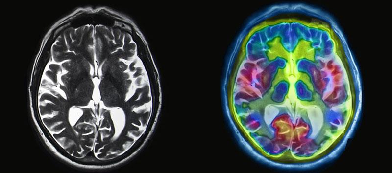
But MRI and CT scans will only show structural damage. So, they’re helpful if you have a severe traumatic brain injury, but if you’ve had a mild traumatic brain injury (mTBI, aka concussion), they likely won’t show anything. If your structural MRI scan comes back as normal, many doctors won’t do much in the way of follow-up.
However, that doesn’t mean you’re out of options. If you’re here, you’ve probably heard of SPECT scans and functional MRI. These imaging tests can show dysfunction resulting from an mTBI.
But which one is right for you? In this post, we’ll explain:
- What SPECT imaging and fMRIs actually are
- What it’s like to get a SPECT scan or fMRI
- What they can show, and what that means for your diagnosis.
If you’re experiencing symptoms that won’t go away after a concussion, we can help. On average, our patients improve by 75% after treatment. To learn about diagnosis and treatment options, schedule a consultation.
Note: Any data relating to brain function mentioned in this post is from our first generation fNCI scans. Gen 1 scans compared activation in various regions of the brain with a control database of healthy brains. Our clinic is now rolling out second-generation fNCI which looks both at the activation of individual brain regions and at the connections between brain regions. Results are interpreted and reported differently for Gen 2 than for Gen 1; reports will not look the same if you come into the clinic for treatment.
All About Brain SPECT Scans

What is a SPECT Scan?
SPECT stands for “single photon emission computed tomography.” That’s a mouthful, so here’s what it translates to:
Patients are injected with a radioactive isotope that emits gamma rays (electromagnetic radiation emitted by a decaying atom). As the isotope (also known as a radioactive tracer) travels through the patient’s bloodstream, it continues to emit radiation. (We know how that sounds, but it really is a safe level of radiation).
A special “gamma camera” is positioned above the patient and able to detect the gamma rays emitted by the isotope. A computer then triangulates in 3-dimensional space where the gamma rays are coming from.
In other words, it looks at multiple data points to figure out where the isotope was in your body when the radiation was emitted.
Over time, collecting multiple “images” can show physicians if blood flow in your body has been impacted by your condition.
Note: You may be more familiar with a PET scan (positron emission tomography). The imaging technique is very similar to SPECT scanning, since both use radioactive tracers to investigate blood flow. PET has greater resolution and fewer image artifacts, but it has other drawbacks.
What Can a SPECT Scan Show?
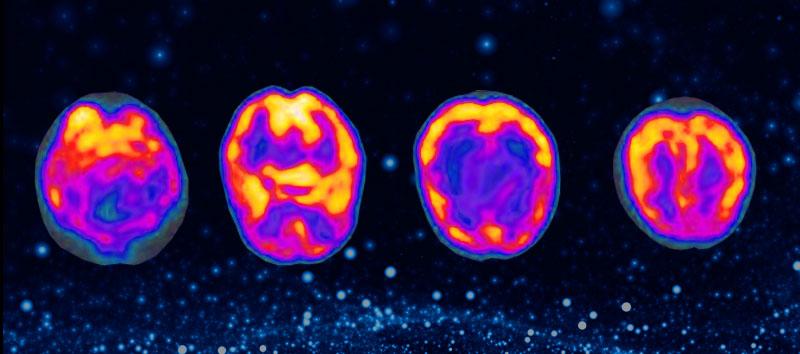
SPECT scans can be used to look at the heart, brain, and a few other things. In the brain, it can be used in evaluating conditions such as neurodegenerative diseases, stroke, seizures, tumors, and other brain trauma. In the heart, it’s most commonly used to view how well the heart is moving the blood that comes through it.
Because of the delay between when the isotope emits gamma rays and when the camera records them, combined with the inaccuracy of having to triangulate their position, SPECT images are an average over time rather than an instantaneous picture. In many cases, it can take 10-15 minutes to get that average image.
Because of that, it can identify certain conditions better than others. If you’ve had a concussion, brain SPECT imaging can confirm whether you have or have not sustained brain dysfunction after the injury. Unfortunately, it often can’t provide more detailed information about specific regions and how they were affected.
How Long Does a SPECT Scan Take?
The time you spend on a SPECT scan depends somewhat on where you’re getting the scan and why you’re getting the scan. They generally range from 1-2 hours (including the time needed for the injection).
The actual scan itself can take as few as 30 minutes.
How Much Does a SPECT Scan Cost?
SPECT scans are one of the more affordable ways to image the brain, but prices can fluctuate a lot based on location, the purpose of the scan, and what additional interpretation is involved.
According to MDsave, brain SPECT scans range from $1,300 to over $3,500.
Does a SPECT Scan Have Any Side Effects?
The main possible side effect of a SPECT scan is having an allergic reaction to the injected isotope. Some people can have bruising and soreness around the site of the injection as well.
If you’re nervous about the radiation, that’s understandable. As far as Western medicine is concerned, however, it’s perfectly harmless. Most people get more radiation from being on a plane at 30,000 feet in the air than they would from a SPECT brain scan.
All About Brain fMRI Scans
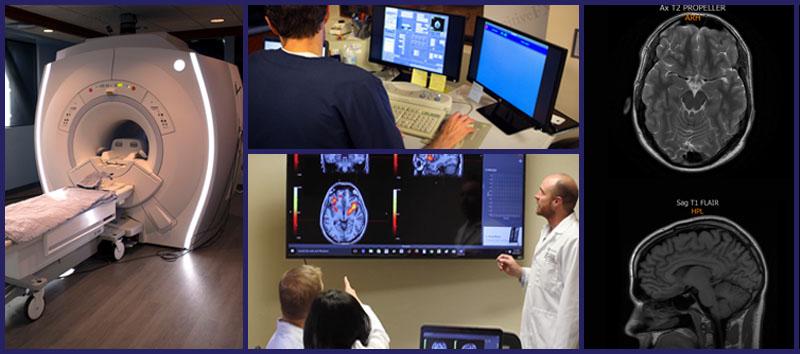
What is an fMRI?
fMRI scans (functional nuclear magnetic resonance imaging) work through a combination of radio waves and magnets. Engineers have figured out how to magnetize soft tissues — such as the brain — very precisely. When you send radio waves through those magnetized tissues, the magnetic field changes the radio wave.
Sensors detect even minimal changes in the radio waves to form a 3D image of the scanned tissue. The only limits to how fine the imaging can become are human ingenuity and engineering skill.
In a structural MRI, that information is used to examine the physical integrity of the brain (or any other organ being imaged). It should show any physical brain damage you’ve sustained. A functional MRI is used to observe blood flow. Since increased cerebral blood flow is tied to increased brain activity, fMRI can show how the brain calls for resources during a given task.
If you’re trying to understand the difference between a structural MRI and a functional MRI, in terms of what it means for patients, this article will help.
What Can an fMRI Show?
fMRIs can show detailed images of blood flow in internal organs. In brain imaging, this means doctors and researchers can see how the brain is managing its oxygen supply and whether the right regions respond in the right way when given a certain task.
For example, we found that patients who have post-concussion syndrome (the condition in which symptoms don’t go away after a concussion) will show tell-tale signs of hyperactivity and hypoactivity in the affected brain regions. Thanks to fMRI, we’re able to pinpoint for post-concussion patients which areas of the brain are dysfunctional and in what way.
To learn more about how we do that, you can read about functional neurocognitive imaging (fNCI), the specific type of fMRI we use.
How Long Does an fMRI Take?
Because fMRI is used more in the research setting than the clinical setting as yet, scan times can vary dramatically. The more you need to know, the longer the scan will take.
At Cognitive FX, an fNCI takes about 45 minutes. During that time, patients take six different cognitive tests while we image their brain to learn how it responds to that stimulation.
How Much Does fMRI Cost?
That depends on who you’re asking — researchers might pay hundreds of dollars per hour for access to one, but if you’re part of a clinical trial, it might be free. That said, if you’re looking to get a brain fMRI for diagnostic purposes, you’ll be charged for both the scan and whatever diagnostic analysis is performed.
At Cognitive FX, charges for an fNCI can run from $3,500 to $5,250, depending on several factors (such as whether you pay in full at the visit or via a payment plan, get the scan as part of a treatment package, etc.).
Does an fMRI Have Any Side Effects?
fMRI does not have any side effects per se, but there are situations in which you might not be able to use it. Some types of foreign metal objects in your body (such as surgical implants, braces, or even permanent eye-liner) may prohibit you from entering the MRI scanner. However, the imaging facility will provide you with full details before you commit to undergoing the scan.
If you have extreme anxiety or fear of enclosed spaces, that would also pose a challenge. fMRI is completed in an enclosed space and is very loud (you are given earplugs, headphones, and cushioning to make the noise more tolerable).
SPECT vs. fMRI: Which is Better?
fMRI is a higher quality test than SPECT, for a few reasons. However, which functional neuroimaging test you need depends on your situation.
The spatial and temporal resolution of fMRI is significantly better: fMRI can see things down to a few millimeters, whereas SPECT resolution is on the centimeter scale.
In other words, fMRI has at least 10x better spatial resolution.
When it comes to temporal (time) resolution, there’s no comparison. SPECT gives an image from 10-15 minutes of activity at a time. fMRI, on the other hand, can give a second-by-second picture of how your brain reacts to given stimuli.
While both methods can show if your brain has been affected by a concussion, fMRI can tell you which parts of your brain were affected (Thalamus? Basal ganglia? Prefrontal cortex?) and how (hyperactive or hypoactive). The latter information is far more useful: If we know which areas of the brain are affected, we can tailor treatment to target those regions. This insight into how your brain function has been impacted by injury is invaluable during treatment.
SPECT makes more sense than fMRI in the case of easier-to-see conditions such as stroke and seizure. Since a SPECT scan is typically cheaper than fMRI, there’s no reason not to use it when it will do the job. But for concussion diagnosis, fMRI provides much more robust, clinically useful data.
fMRI for Concussion Diagnosis
All that being said, it’s important to mention that neither fMRI nor SPECT can be used to diagnose a concussion unless the doctor reading the scans has the right information and tools available.
At Cognitive FX, we do a type of fMRI called fNCI, or functional neurocognitive imaging. It’s what allows us to pinpoint which brain regions were affected (as mentioned above).
fNCI uses the same technology of an fMRI, but the imaging process involves having patients perform standardized tasks while in the MRI machine. Over years of research, we built a database of healthy and unhealthy brains performing these tasks. We know which areas of the brain are supposed to be active during these tasks, and how much or little these areas should respond to each separate task.
When we give a patient an fNCI, we analyze the images of their brain to see which areas of their brain are not working optimally. This process allows for accurate diagnosis of injuries that hinder brain function (such as concussion, but sometimes other conditions like brain dysfunction from carbon monoxide poisoning.)
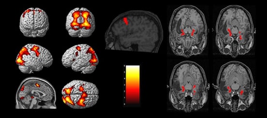
After the test, we meet with patients to discuss their results and how that will affect their treatment. Patients get an overall score and several pages breaking down how each brain region we scanned performed. Here’s an example that one of our patients agreed to share:
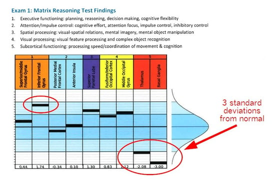
From Olivia’s story: “The fNCI showed that my thalamus was hypoactive and my basal ganglia was completely out to lunch. 3 standard deviations from normal basically means that there was no activation seen in that area on the fMRI. All the work was being routed around it — causing fatigue and stress on the rest of my brain. The inferior frontal gyrus was trending toward hyperactive (using too many resources for given tasks).”
Because fNCI is a kind of fMRI, the test is noninvasive and harmless. There is no radiation, however, the same restrictions around metal apply.
What You Can Do About Concussion Recovery
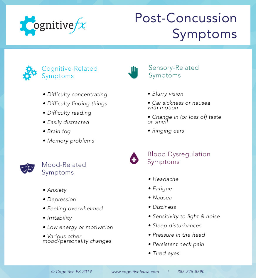
For many patients, concussion symptoms resolve after about two weeks. But for others, those symptoms just won’t seem to go away. If that describes your situation, you may have post-concussion syndrome. We’ve listed some of the symptoms in the chart below:
Your best bet is to seek treatment from a doctor or clinic that specializes in post-concussion treatment. If you’d like to know more about how we can help you, sign up for a consultation.
Or, if you’d like to learn about choosing a clinic, here’s our post on the best concussion clinics in the U.S.
Active Recovery
In the meantime, there are things you can do to improve your chances at a good recovery. Do your best to fit these into every day:
- Plenty of rest (if you’re having difficulty, this post on post-concussion sleep may help).
- Light physical activity for 30 minutes per day (at whatever level you can tolerate without causing symptoms). Here’s our guide to exercising safely after a concussion.
- Cognitive activities like reading or logic puzzles, as tolerated.
- Work or school activities, as tolerated.
Conclusion
Knowing whether you need an fMRI or SPECT scan comes down to a matter of how much you need to know in order to receive effective treatment. If you’re suffering from post-concussion syndrome, a SPECT scan is better than nothing, but an fMRI is significantly more useful than a SPECT scan.
If you’ve been suffering from lingering symptoms after a concussion and haven’t found relief, you’re not imagining things: up to 30% of people who have had a concussion endure lingering symptoms that do not go away without treatment. We’ve written at length about some of the more common issues our patients face, such as headaches, memory problems, relentless fatigue, and more.
To learn more about treatment options and what you can do next, schedule a consultation.