Diagnosing a concussion can be a challenging process. There is no definitive test to confirm the diagnosis with 100% certainty. Instead, doctors often rely on subjective descriptions of symptoms and simple neurological examinations to check systems such as vision, balance, and cognition.
While many doctors order MRI (magnetic resonance imaging) scans or CT (computed or computerized tomography) scans to check the brain for more serious damage, this technology is not suitable for detecting the more subtle changes associated with mild traumatic brain injuries (mTBI or mild TBI).
CT scans are beneficial for finding structural problems such as bone fractures or other injuries that require neurosurgical intervention. For this reason, CT scans are still useful in ruling out other conditions that could result in the same or similar symptoms as a concussion.
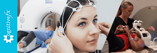
The good news is that for patients struggling with long-term symptoms after a concussion, there is a neuroimaging tool that can actually help with diagnosis: the functional NeuroCognitive Imaging (fNCI) scan. This special type of MRI scan relies on a technology that has been reserved mostly for research purposes, but it has become our method of choice to assess patients that come to our clinic because it allows us to “see” which regions of the brain are dysfunctional post-concussion.
In this post, learn…
If you’re experiencing emotional and physical symptoms that won’t resolve after a concussion, you’re not alone. 95% of our patients experience statistically verified restoration of brain function after treatment. To see if you are eligible for treatment, schedule a consultation.
Note: Any data relating to brain function mentioned in this post is from our first generation fNCI scans. Gen 1 scans compared activation in various regions of the brain with a control database of healthy brains. Our clinic is now rolling out second-generation fNCI which looks both at the activation of individual brain regions and at the connections between brain regions. Results are interpreted and reported differently for Gen 2 than for Gen 1; reports will not look the same if you come into the clinic for treatment.
What is a CT scan?
A CT scan — short for Computed Tomography scan — is a non-invasive diagnostic tool used to detect certain diseases and internal injuries. Over the past 25 years, it has undergone many technical improvements to become one of the most widely used imaging tools today.
In a way, CT scans are like x-rays because they both rely on ionizing radiation to “see” inside the body. However, an x-ray relies on a broad beam of radiation aimed at the body from only one angle. In contrast, a CT scan uses a thin beam to create a series of images taken from multiple angles while revolving around the patient. The information from each angle is fed into a computer, creating a cross-sectional black and white picture representing a slice of a particular area of the body.
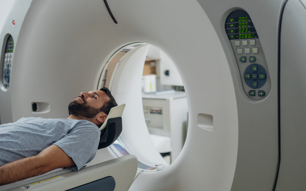
During the scan, you lie down on a flatbed that goes inside the CT scanner, which looks like a giant donut. As the scanner is working, the ring rotates around a small section of your body as you slowly move through it. You will be asked to lie still and follow simple breathing instructions to avoid blurred images.
Once the scan is completed, the computer can layer all the individual slices and create a three-dimensional view of the target organ. Doctors can rotate the 3D image to look at it from different angles. It provides a much more detailed picture than standard x-rays, which are only two-dimensional.
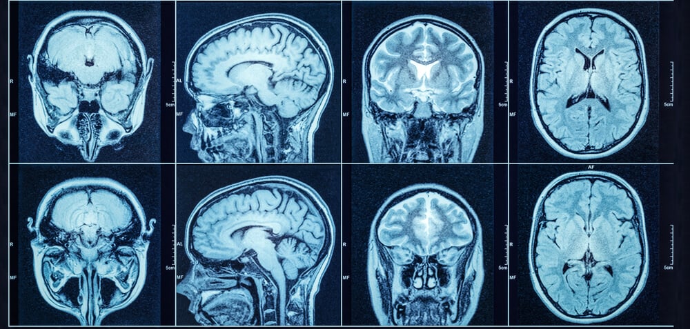
CT scans can identify bone, soft tissue, and blood vessels because they have different densities:
- High-density structures, such as bone, absorb large amounts of radiation, which means a lower amount is detected by the scanner on the opposite side of the body. These show up as white.
- Low-density structures, like internal organs, absorb less radiation, and the scanner detects a greater signal. These show up as various shades of gray.
- Empty spaces don’t absorb any radiation and show up as black.
Risks associated with CT scans
Since CT scans rely on ionizing radiation, you may be concerned about how this exam can increase your risk of developing cancer later in life.
Standard x-rays use relatively low amounts of radiation. The radiation exposure from a head CT scan is slightly higher, but the increase in the risk of developing cancer is still minimal.
If you’re worried, ask your doctor whether the procedure is really necessary and what the risks and benefits are. Pregnant women may want to discuss alternative exams, such as an MRI, to avoid exposing their baby to radiation.
Can CT Scans Be Used to Diagnose a Concussion?
A CT scan is an excellent imaging tool to assess structural damage to bones, soft tissues, and joints — including lesions, fractures, or other abnormalities. It can also detect certain diseases — such as cancer, heart disease, and liver disease — as well as show internal injuries and bleeding.
A CT scan cannot diagnose concussion. That said, the lack of structural findings can help classify your injury as a mild TBI versus moderate to severe (which can be classified with CT findings in addition to other evaluations such as the duration of loss of consciousness).
In most cases, CT scans of concussed patients do not show any abnormal findings, even if the patient is experiencing clear signs of concussion, such as dizziness, nausea, and headaches.
Studies have found that less than 10% of patients with minor head injuries have positive findings on CT, and less than 1% require neurosurgical intervention.
What a CT scan isn't going to tell you, even if you have a moderate to severe TBI, is why your symptoms are not explained by the findings in CT. That’s because traumatic brain injuries, particularly mTBIs, cause functional rather than structural changes in the brain. In other words, they change how the brain functions, but not necessarily how the brain looks.
To understand the difference, imagine the brain as a business. A CT scan will show you structural facts about the business. It can show you that the company building is intact, workers are present, the lights are on, and so forth. What the CT scan can’t show is whether the business is doing well or poorly. The workers could be arguing, the internet connection might be down, or there could be a major slowdown in work, but the CT scan wouldn’t see that. In other words, your brain might be struggling mightily to perform everyday tasks, but unless that problem was caused by a serious structural problem, the CT scan can’t see that.
Since the signs and symptoms of a concussion are not connected to any damage to the brain detectable by a CT scan, it’s not a useful tool for detecting a concussion.
If you're looking for a way to assess your symptoms right now, our free concussion quiz is based on the same clinical tools (PCSS, SCAT6) used in professional sports medicine.
When Doctors Use CT Scans After a Concussion

Despite its inability to diagnose concussions, doctors still use CT scans to diagnose severe conditions that may happen as a consequence of head trauma. Most emergency department doctors will recommend a CT scan for patients with:
- Loss of consciousness for more than 5 minutes after the injury
- Evident skull injuries
- A Glasgow Coma Scale score below 14
- Worsening or severe headache and multiple episodes of vomiting
- Severe amnesia
- One pupil larger than the other
- Fluid or blood coming out from ear or nose
- Alcohol or drug intoxication
- Confusion or aggressive behavior
- Very young children or over the age of 60
For these patients, one of the major worries is an intracranial hemorrhage, more commonly known as bleeding in the brain. This type of bleeding puts pressure on the affected area and restricts blood flow, ultimately affecting how the brain functions.
These patients may need surgical intervention, and CT scans play a crucial role in identifying, following, and managing this condition. Accumulated blood is exceptionally dense compared to brain tissue, and brain bleeds are usually not difficult to diagnose with a CT scan.
A second condition that can be detected with a CT scan involves skull fractures. In the image, fractures will appear as lines in the bone, often accompanied by blood in the surrounding areas. Depending on the location, size, and type of fracture, these may need to be surgically repaired to relieve pressure or to stop bleeding. Although an x-ray of the skull may detect fractures, CT is normally the imaging tool of choice for most doctors.
With the rising occurrence of injuries caused by firearms, it is becoming common to find foreign bodies, such as bullets, lodged inside the brain. In addition to finding these foreign objects, CT scans can help track any movements and anticipate further complications.
We can help certain patients recover from the long-term symptoms of traumatic brain injury (TBI). Reach out to our team to schedule a consultation to see if you meet our treatment criteria.
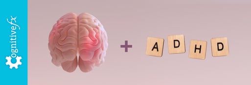
What About Diagnosing Post-Concussion Syndrome (PCS)?

Post Concussion Syndrome (PCS) occurs when patients still have lingering symptoms months or even years after the initial head injury. Most people experience symptoms for the first 7 to 10 days after a concussion, then recover within a few weeks. But for up to 30% of concussion patients, symptoms just won’t resolve on their own.
Patients with PCS often struggle to obtain a diagnosis as many doctors are not well educated on the topic. But, why is this diagnosis so difficult? The brain controls or influences most mechanisms in the body, so symptoms can appear anywhere, from your sleep cycle to your vision. As a consequence, symptoms are often attributed to something other than the original concussion and treated individually.
Unfortunately, a CT scan cannot confirm a PCS diagnosis. In fact, a study found only two abnormal imaging tests (including x-rays, MRIs, and CTs) out of 32 examinations in PCS patients, neither of which actually confirmed a PCS diagnosis or altered how the patient was treated. After a cost of more than $129,000 to find only two abnormal scans, the authors (unsurprisingly) concluded that using CT scans to diagnose PCS was ineffective and costly.
Alternative diagnostic tests like Positron Emission Tomography (PET) or Single-Photon Emission Computerized Tomography (SPECT) scans have also failed to provide actionable intelligence for PCS patients.
But one technique stands out amongst the rest: functional MRI (fMRI).
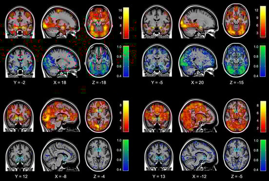 Imaging studies taken from Hariri et al.
An fMRI uses the same technology as a standard MRI, but these two methods produce very different results. MRIs are used to assess brain structure, whereas fMRIs can detect changes in blood flow or blood oxygen levels to produce a functional image of the brain.
In other words, MRIs produce static images of the brain, while fMRIs produce “active” images of what is going on inside the brain while it’s working.
For example, when an area of the brain is more active, blood will flow to the area and appear as a hot signal on the fMRI. In contrast, if an area of the brain is less active, it will show a weaker signal.
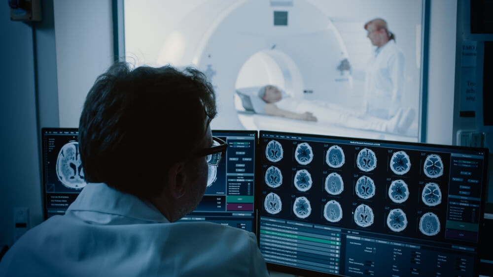
At Cognitive FX, we combine fMRI with a specific protocol of cognitive tasks, then compare each patient’s scans with a database of brain scans from healthy and unhealthy patients. As a result, we obtain the standard deviation from normal for dozens of regions of the brain in addition to communication between regions. That way, we can not only tell which patients have post-concussion syndrome, we can also know which regions of the brain are dysfunctional.
This approach is known as a functional NeuroCognitive Imaging (fNCI) scan and is the imaging tool of choice at Cognitive FX.
fNCI is over 98% accurate at diagnosing post-concussion syndrome.
How Does fNCI Produce a Concussion Diagnosis?
As described earlier, this scan of the brain measures the increase or decrease of blood in different brain regions. By having patients perform six carefully designed cognitive tasks during the scan, we can see how specific brain areas react when they’re “called” to work.
The results show which parts of the brain react normally and which parts are not using blood flow efficiently.
In dysfunctional regions, the brain is not getting the right amount of oxygen and nutrients at the right time. Patients might struggle to complete the task that requires the use of that part of the brain, or completing it might tire them far more quickly than it should. This dysfunction leads to cognitive deficits and physical symptoms.
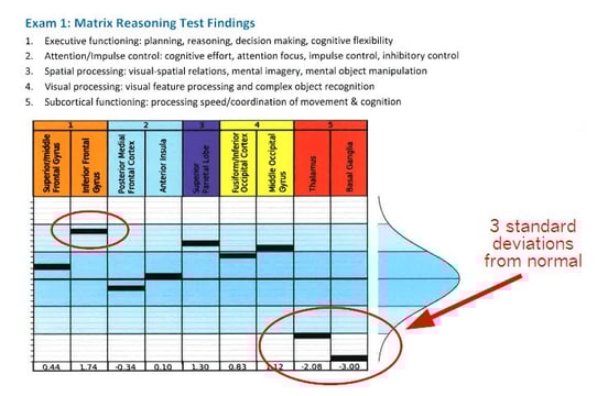 One of our patients explained her fNCI results in her post-concussion syndrome recovery story: “The fNCI showed that my thalamus was hypoactive and my basal ganglia was completely out to lunch. 3 standard deviations from normal basically means that there was no activation seen in that area on the fMRI. All the work was being routed around it — causing fatigue and stress on the rest of my brain.” Note that this was from an older version of fNCI; your scan may look different.
Under normal circumstances, nerve cells in the brain receive a dynamic supply of oxygen and nutrients from the surrounding blood vessels. This connection is called neurovascular coupling (NVC). NVC makes sure blood flows to the areas where it's needed, when it’s needed, to ensure brain cells function normally. For example, if you’re reading a book, there’s an extra boost of blood sent to the visual and language regions so that they can do their job.
After a concussion, however, the connection between brain cells and the blood vessels that supply them with nutrients and oxygen is disrupted, making it difficult for certain parts of the brain to work correctly. This is known as neurovascular coupling dysfunction and is one of the root causes of concussion symptoms.
Since fNCI can provide information about how blood flows to different regions of your brain, it can detect this dysfunctional communication between nerve cells and blood vessels that is characteristic of PCS.
Treatment at Cognitive FX
After your fNCI scan, we meet with you to review your results and discuss your customized treatment plan.
At our clinic, we combine aerobic exercise with a range of multidisciplinary therapies, including neuromuscular therapy, occupational therapy, cognitive therapy, neurointegration therapy, sensorimotor therapy, vestibular therapy, and more. Alternating therapy with physical activity makes treatment more effective.
During each day of our treatment, patients see different therapists to receive their prescribed treatments. Our therapists know how to adapt each exercise according to each patient’s needs. They leverage information from the initial fNCI scan to create the most effective treatment sessions.
During cognitive therapy, for example, patients have to complete a series of exercises addressing cognitive issues — such as processing speed, attention difficulties, sensitivity to noise, memory problems, and more.
For vision and vestibular therapy, patients participate in exercises to address visual problems, including sensitivity to light, vertigo, balance challenges, and more.
Neuromuscular therapy is designed to address the most common physical symptoms of concussion, including headaches, dizziness, and nausea. The aim is to retrain the body to work better in conjunction with the brain.
In addition to these and other types of therapy, patients also attend two sessions with a psychologist to help address any emotional difficulties caused by the brain injury.
At the end of treatment, each patient does another fNCI scan to see how much their brain function has changed. For most patients, the improvement they’ve already felt as symptoms start to dissipate is reflected in the positive changes observed in the second scan. On average, we see improvements of over 75% after therapy at our clinic.
We also meet again with each patient to arrange for a post-care plan. This incorporates recommendations from our therapists for a range of exercises to do at home to ensure that patients continue to improve after their time with us.
If you’re experiencing emotional and physical symptoms that won’t resolve after a concussion, you’re not alone. 95% of our patients experience statistically verified restoration of brain function after treatment. To see if you are eligible for treatment, schedule a consultation.


.png?height=175&name=Why%20Post-Concussion%20Syndrome%20Causes%20Tingling%20Hands%20(And%20What%20to%20Do).png)